Tendon Diagram : Tendon Vs Ligament Medlineplus Medical Encyclopedia Image : The tendon is firmly connected to muscle fibres at one end and to components of the bone at its other end.
Tendon Diagram : Tendon Vs Ligament Medlineplus Medical Encyclopedia Image : The tendon is firmly connected to muscle fibres at one end and to components of the bone at its other end.. To be connected together by the joints, some bones of the. Its muscle belly is in the forearm. Tendons, located at each end of a muscle, attach muscle to bone. Ligaments and tendons are adapted in response to changes in mechanical stiffness. Groin strain treatment rehabilitation exercises although there is often swelling oedema as a result of a groin strain this is often not visible to the eye groin strains are graded 1 2 or 3 depending on the extent of the injury groin muscle diagram diagram muscles in groin area male groin muscle diagram diagram muscles in groin area male anatomy groin human photo groin.
Flexor tendon lacerations are classified into five zones 2, 15, 16. Again, our knowledge of how mechanical stimulus mediates ligament and tendon structure is more empirical and less. Tendons are remarkably strong, having one of the highest tensile strengths found among soft tissues. The hand incorporates countless muscles, bones, tendons and ligaments into simple motion and this chart covers them all. The fcu tendon is one of two tendons that bend the wrist.
Tendons High Res Stock Images Shutterstock from image.shutterstock.com Allows the action of raising the foot. The largest of these shoulder muscles is the. Lower back muscle diagram anatomy does degenerative disc disease affect the lower back muscle? Flexor tendon lacerations are classified into five zones 2, 15, 16. Ligaments and tendons are adapted in response to changes in mechanical stiffness. Groin strain treatment rehabilitation exercises although there is often swelling oedema as a result of a groin strain this is often not visible to the eye groin strains are graded 1 2 or 3 depending on the extent of the injury groin muscle diagram diagram muscles in groin area male groin muscle diagram diagram muscles in groin area male anatomy groin human photo groin. A tendon is a band of tissue that connects a muscle to a bone. The tendon is firmly connected to muscle fibres at one end and to components of the bone at its other end.
Medical labeled diagram closeup with muscle, transverse carpal ligament, median nerve, tendon sheath, flextor tendons and bones.
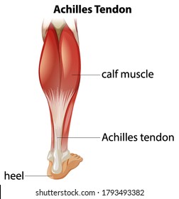
Intermediate back muscles and c. The largest of these shoulder muscles is the. Tendons, located at each end of a muscle, attach muscle to bone. The achilles tendon attaches the muscles of the calves to the bones of the ankle and foot. Related posts of shoulder muscles and tendons diagram. Medical illustration of human arm muscles, veins and nerves. The two peroneal tendons in the foot run side by side behind the outer ankle bone. Foot anatomy diagram, foot joint diagram, foot sprain diagram, foot tendons and ligaments pain, leg tendon diagram, peroneal tendonitis, foot, foot anatomy diagram, foot joint diagram, foot sprain diagram, foot tendons and ligaments pain, leg tendon diagram, peroneal tendonitis. In the back and elsewhere in the body, tendons attach muscles to bones. This muscle diagram is interactive: The joint is strengthened and stabilized by adjacent muscles and tendons, especially by the musculotendinous rotator cuff. Tendons are the connection between bones and muscles. Jul 05, 2018 · the foot diagram has a complex structure made up of bones, ligaments, muscles, and tendons.
The bones together make up the hip. On the other hand, the insertion is where a tendon attaches that muscle to the *more* movable bone. The anterior cruciate ligament prevents the femur from sliding backward on the tibia (or the tibia sliding forward on the femur). Lower back muscle diagram anatomy does degenerative disc disease affect the lower back muscle? The achilles tendon is the largest.
Knee Human Body Anatomy Tendon Patellar Ligament Others Human Human Back Arm Png Pngwing from w7.pngwing.com A tendon is a band of tissue that connects a muscle to a bone. A muscle's origin is where a tendon attaches it to the *less* movable bone. Its muscle belly is in the forearm. This diagram depicts muscle in the body 744×1054 with parts and labels. Ligaments and tendons are adapted in response to changes in mechanical stiffness. Allows the foot to be turned inward and also supports the arch of the foot. Diagram depicting the bones, ligaments and muscles throughout the hand and fingers. The hip itself is a ball and socket joint, much like the shoulder.the structures necessary to create this joint are the socket, the joint capsule, muscle, ligaments, and the neck.
A diagram depicting the muscles in the upper body, from the backside.
The rotator cuff is a group of four muscles and tendons that surround the glenohumeral joint. Tendon diagram diagram illustrating tendonitis and tendon rupture symptoms can vary from aches or pains and local joint stiffness, to a burning that surrounds the whole joint around the inflamed tendon. The pubis, ischium, and ilium together constitute the pelvis while the thigh bone is the femur. Related posts of shoulder muscles and tendons diagram. The fcu tendon is one of two tendons that bend the wrist. Tendon diagrams and design force vectors. On the other hand, the insertion is where a tendon attaches that muscle to the *more* movable bone. This diagram depicts muscle in the body 744×1054 with parts and labels. When the muscles tighten (contract) arguably, the most important tendon is the achilles tendon, which allows the calf muscles to move. The achilles tendon is the largest. The biceps muscle has two tendon attachments. Muscles of the shoulder : There are over two dozen gorgeous and painstakingly detailed illustrations on this chart, from the extensor pollicis longus to the flexor digitorum.
The achilles tendon enables us to walk, without it we would not be able to raise our heels of the ground. The ecu tendon works along with the ecrl and ecrb to straighten the wrist. Medical labeled diagram closeup with muscle, transverse carpal ligament, median nerve, tendon sheath, flextor tendons and bones. The achilles tendon is also called the calcaneal tendon. The achilles tendon is the largest.
14 034 Tendon Stock Photos Pictures Royalty Free Images Istock from media.istockphoto.com Jul 05, 2018 · the foot diagram has a complex structure made up of bones, ligaments, muscles, and tendons. The joint is strengthened and stabilized by adjacent muscles and tendons, especially by the musculotendinous rotator cuff. The achilles tendon is the largest. Groin strain treatment rehabilitation exercises although there is often swelling oedema as a result of a groin strain this is often not visible to the eye groin strains are graded 1 2 or 3 depending on the extent of the injury groin muscle diagram diagram muscles in groin area male groin muscle diagram diagram muscles in groin area male anatomy groin human photo groin. Tendon diagrams and design force vectors. The achilles tendon enables us to walk, without it we would not be able to raise our heels of the ground. Learn about these muscles, their origin and insertion points, and their functional anatomy. Attaches the calf muscles to the calcaneus, most important muscles for running, jumping, walking etc.
Your biceps tendons attach the biceps muscle to bones in the shoulder and in the elbow.
The rotator cuff is a group of four muscles and tendons that surround the glenohumeral joint. The tendon runs down the back of your lower leg from the back of the knee to the heel. Attaches the calf muscles to the calcaneus, most important muscles for running, jumping, walking etc. There are over two dozen gorgeous and painstakingly detailed illustrations on this chart, from the extensor pollicis longus to the flexor digitorum. The achilles tendon enables us to walk, without it we would not be able to raise our heels of the ground. Ligaments join the knee bones and provide stability to the knee: Tendons attach muscles to bones. Foot anatomy diagram, foot joint diagram, foot sprain diagram, foot tendons and ligaments pain, leg tendon diagram. The hand incorporates countless muscles, bones, tendons and ligaments into simple motion and this chart covers them all. This tendon connects the patella (kneecap) to the tibia. The achilles tendon is the largest. Muscles of the shoulder : The achilles tendon is also called the calcaneal tendon.


0 Komentar